

 Menu
Menu

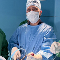
Dr. Bijay Kumar Sahu
Orthopaedic Surgeon
Questions? We’re here to help! Fill the form or chat with us now.

Advanced Shoulder, Knee & Upper Limb Care with Dr. Bijay Kumar Sahu
A total shoulder replacement is an open procedure that involves replacing the shoulder joint with a specially designed prosthesis.
A total shoulder replacement may be recommended for patients suffering severe osteoarthritis in the glenohumeral joint.
A total shoulder replacement is an open procedure that involves replacing the shoulder joint with a specially designed prosthesis. The prosthesis consists of two components. The first is a humeral head component, a metal ball secured into the humeral bone with a stem.The other is a glenoid component, which replaces the glenoid and allows smooth movement of the new humeral head.
The system closely replicates the shoulder joint and relieves pain and discomfort by replacing the severely affected joint. The wound is generally closed with absorbable sutures. However, instructions will be given by Dr Bijay Kumar Sahu post-surgery as to wound and dressing procedures.
Following surgery, an X-Ray and a CT scan will be performed on day 1 post-op, to check that the position of the prosthesis is satisfactory. Physiotherapy will be commenced on day 1 post-op and patients will be given a rehab programme by their physiotherapist.
Following an anatomic total shoulder replacement, a sling will be worn post-op for 6 weeks following the surgery.
Following a reverse total shoulder replacement, the length of time a sling is required depends on the details of the operation. For instance, if there has been no subscapularis repair at the time of the reverse shoulder replacement, then a sling may be recommended only for a couple of weeks. If a latissimus dorsi transfer is required at the time of a reverse total shoulder replacement a sling or a brace will be required to be worn full-time for 6 weeks.
A reverse total shoulder replacement may be considered inpatients suffering severe joint arthritis with irreparable rotator cuff tears or patients with a deficient rotator cuff suffering superior migration of the humeral head. Another consideration for a reverse total shoulder replacement may include a mal-union of a proximal humeral fracture or an irreparable proximal humeral fracture.
The prosthesis used in a reverse total shoulder replacement is like a total shoulder replacement, however, the prosthesis is reversed.Instead of the ball on the end of your humerus, a socket is placed and instead of the socket on your shoulder blade (glenoid) a ball, or glenosphere, is placed. Hence the socket and the ball are reversed.
If there is severe weakness in the external rotation of the arm or complete irreparable tears of the rotator cuff, a latissimus dorsi transfer may be used to improve the external rotation. This involves transferring the latissimus dorsi to act as an external rotator, rather than an internal rotator of the humerus. This provides stronger support and stability for the new prosthesis and a greater active range of motion.
On day 1 post-operatively an X-Ray and CT scan will be performed to check the position of the prosthesis and physiotherapy will commence. Patients will be in a sling for up to 6 weeks following surgery. The exact time in the sling is determined by the specific details of the surgery.
Many patients are only in a sling for a few weeks post-op.
A review with Dr Bijay Kumar Sahu will be organised at 6 weeks post-op. Usually, patients are reviewed at 6 weeks, 12 weeks, 6 months, 12 months and 24 months post-op. Final check scans are performed at 12 months post-op.
I use the following prostheses in most instances:
Wright, Tornier Ascend Flex
Zimmer Biomet Comprehensive
These prostheses have established track records in the Australian Shoulder Registry. With excellent performance in terms of longevity.
An acromioplasty involves shaving the under-surface of the acromion. The acromion is a projection of bone extending from the shoulder blade, over the top of the shoulder joint and provides attachment for muscles around the shoulder including the trapezius and deltoid muscles.
An acromioplasty is typically performed for patients where their rotator cuff is pinching on the under-surface of the acromion and the coracoacromial ligament.
Keyhole surgery is used to shave the under-surface of the acromion to provide greater room for the rotator cuff tendons to fit under the acromion. If enough room can be created, then the rotator cuff tendons may not pinch on the under-surface of the acromion and the pain of impingement can be completely relieved.
Patients with calcific tendinitis and large calcium lumps can be treated with an arthroscopic excision of the calcium deposit. This may also be performed in conjunction with an acromioplasty.
The region of the tendon with the calcium inside it can usually be identified by the visualisation of an inflamed area of the tendon. The location can be confirmed by probing the tendon with a needle until calcium is seen in the tip of the needle. Once the calcium is located a small longitudinal incision is made in the tendon in line with its fibres and the calcium is removed with an arthroscopic shaver and curette.
If the rotator cuff tendons have been torn off their attachment to the bone on the humerus, then a repair may be required.
This surgery can be performed as an open or arthroscopic repair. Arthroscopic surgery involves the latest techniques for rotator cuff repair and is as successful or more so than open rotator cuff repair. The arthroscopic technique avoids the need for a large incision and the post-operative pain and discomfort following the arthroscopic procedure is consequently considerably less than the open procedure. The need for an open surgery scar is also avoided.
Patients suffering severe irreparable tears of the posterosuperior rotator cuff may be considered for a latissimus(lat) dorsi transfer. The posterosuperior tendons of the rotator including the infraspinatus and the posterior supraspinatus tendons act as external rotators for the shoulder. These tendons assist movement including lifting and use of the arm overhead.
A lat dorsi transfer is a procedure that involves transferring the lat dorsi tendon and possibly the teres major tendon from an internal rotator to an external rotator. This procedure is commonly used in conjunction with a reverse total shoulder replacement.
Dr Bijay Kumar Sahu is currently developing an all-arthroscopic method of performing a latissimus dorsi transfer. He has recently successfully performed his first arthroscopic lat dorsi transfer.
Tendonitis means inflammation of the tendon. The rotator cuff tendons are particularly prone to tendonitis, and the one that is most often involved is the supraspinatus tendon.
The Arthroscopic Bankart Repair is an effective procedure to treat patients that have anterior shoulder instability. Many patients who suffer a traumatic anterior dislocation of their shoulder will tear the fibrocartilage labrum at the front of the shoulder. Many of these patients will go on to develop recurrent instability in their shoulder and keep dislocating.
This will have a significant effect on the ability to participate in sports and sometimes also their work. It is the tear in the labrum that is largely responsible for allowing their shoulder to continue to dislocate.
It has been established that if only patients with a pure labral tear are treated with an arthroscopic Bankart repair then the results are as high as an open repair. The aim of surgery is to return people to full normal sporting and work activities and the risk of a re-dislocation in this situation is less than 5% with a well-performed arthroscopic procedure.
The Arthroscopic Bankart procedure repairs this tear in the labrum and by doing so restores stability to the shoulder. This procedure can be performed either open or arthroscopically. Previously it was believed that the arthroscopic repair was not a successful as the open repair.
Patients that have torn not just the labral cartilage at the front of the shoulder but have also chipped off a segment of bone when they dislocated need a surgical procedure that will deal with the loss of bone. My preference in this situation is an arthroscopic Latarjet procedure.
Arthroscopic Brachial Plexus Exploration is undertaken when there is an indication that pressure may be unduly compromising a nerve around the shoulder.
This patient had pain in his shoulder following a shoulder procedure. Arthroscopic exploration allowed Dr Bijay Kumar Sahu to identify a fascial band that was compressing the axillary nerve when the arm was raised.
Dr Bijay Kumar Sahu was able to divide the fascial band and in so doing relieve the patient’s symptoms.
Following non-operative treatment for an arthritic AC joint, an AC joint resection may be discussed. This procedure is done as a keyhole operation and involves removing a segment of bone at the end of the clavicle (collarbone).
Resection of a painful AC joint is very effective in relieving pain. The resected AC joint is replaced by fibrous scar tissue that takes the place of the worn-out, inflamed joint.
In the past, there was a tendency to take bone only off the clavicle in order to excise the AC joint. It has since become apparent that some of these patients have excessive instability symptoms, especially in an anteroposterior direction due to compromising the posterosuperior capsular ligaments.
The Arthroscopic resection allows these important posterosuperior ligaments of the AC joint to be preserved avoiding problems with instability. Open surgery will always involve the division of these important structures which then need to be repaired once the surgery is completed.
Dr Bijay Kumar Sahu now removes approximately 5mm of bone from the distal clavicle and a small amount of bone from the medial acromion. Doing this greatly helps to avoid instability symptoms in his patients.
Acromioclavicular joint (ACJ)dislocations commonly occur following a fall or a blow to the point of the shoulder and are a common sporting injury. A prominent lump will usually be present on the point of the shoulder.
ACJ injuries are graded according to the Rockwood classification system into grades I to VI.
Low-grade type I and II injuries involvecapsular sprain injuries without rupture of the critical coracoclavicular ligaments.
Grade III to VI injuries denote injuries in which the coracoclavicular and acromioclavicular ligaments have ruptured and the clavicle displaces upwards giving the characteristic appearance of the lump on the point of the shoulder.
Grade IV to VI injuries are high-grade injuries and surgical reconstruction is usually recommended.
Grade III injuries, in certain circumstances, are treated with surgical reconstruction.
Dr Bijay Kumar Sahu performs reconstruction of acute AC joint injuries using an all-arthroscopic technique. He has recently had an article describing this all-arthroscopic technique accepted for publication by the prestigious international medical journal the Journal of Shoulder and ElbowSurgery in their techniques in shoulder and elbow surgery.
Arthroscopic repairs of rotator cuff tears are associated with a lower complication rate than that for open repairs. However, both techniques are subject to similar possible complications. Shoulder surgery in Australia is usually performed under general anaesthesia. There are risks associated with any general anaesthetic.
Infection is also a complication that can occur following a rotator cuff repair. The rate of infection is lower following an arthroscopic repair than for an open repair. If infection occurs it can lead to failure of the rotator cuff. Further surgery can be required along with antibiotic treatment to control any possible infection. The rate of infection for an arthroscopic repair is less than 0.2%.
Nerve injury is an extremely rare and serious complication of shoulder surgery. A frozen shoulder can occur following any surgical procedure or any injury to the shoulder.
Frozen shoulder is a condition peculiar to the shoulder. It is one that recovers of its own accord, however, until it does the shoulder is painful and stiff. It can frequently take 12 months or more for a frozen shoulder to resolve after an operation. Treatment for a frozen shoulder is available. Treatment is undertaken for the symptoms of a frozen shoulder and usually includes steroid injections and in severe cases a surgical release of the shoulder.
When a patient is diagnosed with articular cartilage damage, or a defect, it is important to address the injury and treat the condition so that further progression does not occur. Articular cartilage is a very important component of a healthy knee joint. It is a fibrous substance that covers the ends of the bones and is what allows for a fluid, smooth, painless motion of the knee. When a patient has articular cartilage damage, either from a previous injury, overuse, or trauma to the knee, they will experience pain, swelling, and periods of weakness in the knee. Over time, this process will continue to progress and eventually lead to a complete loss of cartilage down to the bone. When this happens, a diseases known as osteoarthritis becomes present. This continues to cause progressive symptoms of knee pain and stiffness.
Osteochondral Autograft Transfers
Osteoarticular Allografts
Microfracture Surgery of the Knee
Knee ligament injuries are a common occurrence among athletes, however, everyday individuals are also at risk for injuring one or more of their ligaments just by doing everyday activities and functions such as running, jumping, and climbing stairs. Knee ligament injuries can affect the ACL (anterior cruciate ligament), PCL (posterior cruciate ligament), MCL (medial collateral ligament) or LCL (lateral collateral ligament). A knee ligament injury can be isolated to one specific ligament, or it can be a more complex, multi-ligament injury where more than one ligament is involved. In many cases, when multiple ligaments are affected, other issues with surrounding tissue, muscle, and bone can arise.
Following a thorough physical examination, Dr. Bijay Kumar Sahu will determine if an isolated ligament injury is present, or if multiple ligaments are involved. He is skilled at performing ligament reconstruction.
ACL Reconstruction
Revision ACL Reconstruction
LCL Reconstruction
PCL Reconstruction
MCL Reconstruction
Complex Knee Surgery
Posterolateral Reconstruction
Biplanar Osteotomy
A meniscus injury is common among athletes and can lead to ongoing pain and knee instability. The menisci are c-shaped pieces of fibrocartilage that cover the knee and play an extremely important role in distributing the load across the knee and to protect the articular cartilage. The meniscus is made up of the medial and lateral meniscus each serving as a shock absorber within the knee.
Injuries to both the medial and lateral meniscus will vary in severity and some cases can be considered quite complex and severe. Dr. Bijay Kumar Sahu offers multiple meniscus treatment options and is a skilled expert in treating meniscal injuries. The treatments he offers are listed below.
Discoid Meniscus Surgery
Meniscus Transplant
Meniscus Surgery
Medial Meniscus Root Repair: Post-surgical Recovery Process, Expectations, and Timelines
Meniscus Root Repair
Partial Meniscectomy
When a patient has been diagnosed with osteoarthritis of the knee, the first step in treatment is to try and determine ways to avoid those activities that cause the symptoms. If surgery is the recommended course of action, Dr. Bijay Kumar Sahu will discuss each technique and offer a treatment solution based upon the progression of the disease.
While one thinks about total joint replacement as a common treatment of osteoarthritis, most patients with arthritis can put off joint replacement or avoid it altogether by undergoing other arthroscopic joint preservation techniques. Osteoarthritis treatment can be performed arthroscopically in many patients to clean out the irritated joint lining, to trim out any areas of a meniscal tear, to smooth off the cartilage surfaces, and to remove any bone spurs that may be limiting knee motion. Dr. Bijay Kumar Sahu in an expert in the following osteoarthritis treatment:
Knee Osteotomy
Joint Preservation Surgery
Total Knee Replacement Surgery
Patellofemoral joint injuries are common musculoskeletal issues that can affect individuals of all ages. They represent the most diagnosed injuries among sports medicine specialists and can result from a myriad of causes—most common are sports related activities, overuse, and trauma to the kneecap.
These injuries are described as pain underneath the kneecap, but the symptoms can also be felt on the anterior or posterior sides. Dr. Bijay Kumar Sahu is an expert in treating patellofemoral pain and offers a number of techniques for patients with this problematic knee condition:
Lateral Patellotibial Ligament Reconstruction
Trochleoplasty
Tibial Tubrical Osteotomy
Dr. Bijay Kumar Sahu
Orthopaedic Surgeon
Questions? We’re here to help! Fill the form or chat with us now.

 Close
Close
